

Authors: Murphy, M P.
Figure 1: Magnetic resonance imaging of the left knee demonstrating common medical imaging findings that are associated with meniscal tears (1).
Abstract
In a retrospective study published by the BMJ in 2007, it was reported that knee injuries accounted for 46% of all lower body injuries in the Adult World Hockey Championships & Winter Olympics.
Of those forty-six percent, it was reported that 14.5% of these injuries accounted for meniscal tears and were more prevalent than ACL ruptures (2).
Therefore, the aim of this manuscript is to offer sports medicine insight on how to clinically manage the post-meniscectomy athlete, discuss relevant anatomy, introduce pathological classifications, and offer a hypothesis on how to proficiently manage patient outcomes via PT modalities. These hypotheses are supported by clinical practice guidelines and credible scholarly articles.
Introduction
As a joint, the knee is compromised four bony articulations. These are the distal femur, the proximal tibia, the patella, and fibula. The tibiofemoral joint is classified anatomically as a synovial condyloid joint with two main degrees of freedom. These kinematical degrees of freedom include flexion and extension in the sagittal plane & internal and external rotation in the horizontal plane.

Figure 2: Human anatomy of the tibiofemoral joint from Gray’s anatomical atlas (3).
In regard to anatomical structure, the knee can be divided into two compartments.
Within the internal compartment, exists fibrocartilaginous structures such as the medial and lateral meniscus, which function as shock absorbing tissue, and theoretically prevent bone degradation between the distal head of the femur and the proximal tibial. In addition, ligamentous tissue such as the anterior and posterior cruciate ligament, also reside within the internal compartment, and function to check anterior and posterior glide of tibio-femoral joint
Within the external compartment there exists two more important ligamentous structures that check for valgus or varus kinematic motion. These structures are known as the medial and lateral collateral ligament respectively. Lastly, the popliteal artery and vein vascularize the posterior aspect of the knee, while the genicular artery and vein vascularize the anterior aspect of the knee.
In the realm of sports medicine, the knee is commonly injured as a result of direct physical trauma and is known in this culture as going knee to knee. When this occurs, a valgus or varus STRESS force is often applied to the tibial-femoral joint resulting in compressive and tensile STRESSES being applied to the internal compartments of the knee. As a result, damage can ensue like that shown in figure 3.
The classification system used to diagnose root tears of the meniscus is referred to as the LaPadre system and was first developed in 2015 by MD Robert LaPadre who is an orthopedic knee surgeon from Minnesota.

Figure 3: A visual representation of the LaPrade classification for meniscus root tears of cadaveric specimens. (1).
| Type | Description |
| 1 | Meniscal tear 0 to 9 mm from root attachment that is partially stable |
| 2 | Comple radial tear of the meniscus |
| 3 | Bucket-handle tear of the meniscal root with detachment |
| 4 | Complex oblique tear that extends into the attachment site. |
| 5 | Avulsion fractue of the meniscal root attachment. |
Table 1 – A description of anterior and posterior tears of the medial and lateral meniscal root tears (5).
In order to conduct surgical repairment of the meniscus, an arthroscopic cannula is inserted within the superolateral or superomedial aspect of the suprapatellar pouch and fragments of the fibrocartilage are removed in order to provide a smooth articular surface between the tibia and femur. It has been reported that this procedure takes approximately one hour to complete and is associated with a financial cost of $12,000.
Post surgery, patients are commonly prescribed pain killers, assisted walking devices like crutches, and physical therapy regiments in order to manage pain, build muscular strength, and restore range of motion and coordination.
Within the remainder of this manuscript, we will explore PT modalities that are supported by empirical evidence to manage outcomes of the post-meniscectomy athlete.
Body
According to clinical practice guidelines put forth by Wexner Medical Center, a subsidiary of Ohio State University it is stated that patients with “standard meniscus repair should be routinely monitored for signs and symptoms of DVT’s immediately following surgery” (4).
Once the acute phase of healing has been reached, post-surgery, it is imperative that individuals begin physical rehabilitation to restore functionality of the tibiofemoral joint. Current clinical practice guidelines “high level activity” (4) athletes suggest that these individuals should being rehabilitation with isometric, isokinetic and plyometric training.
It is imperative that, at all stages of clinical management, the practitioner should be aware of the traffic light system. A red light is associated with symptoms that should limit physical activity. A yellow light causes no change in patient outcomes. A green light, demonstrate clinical progression of rehabilitation process.
Within the section below, the reader may view a hypothetical treatment plan for the post-surgical meniscectomy athlete, which is broken down into four stages.
Stage 1 – Pre-Surgery
- Isometric Contractions of the Quadriceps
- TENS Application: for pain control
- Alternating Heat and Cold Pack application
Goal(s): During this phase of rehabilitation, it is the goal to maintain as much muscle mass within the lower extremity as possible. Following surgery, atrophy due to immobilization is likely to occur. In addition, the TENS unit will be utilized to decrease patient’s perception of pain. Lastly, the alternating use of hot and cold packs will increase vascular flow and allow innate healing processes to be maximized. These modalities should be completed every other day until surgery occurs.
Stage 2 – Weeks 0-2 Post Surgery
- Ultrasound application
- Isometric Contractions of the Quadriceps with NMES : 3 sets of 15 repetitions 3 times per week.
- Patellar Mobilization: Preformed in superior & Inferior glide as well as medial and lateral glide.
- Alternating Heat and Cold Pack application
Goal(s): During this phase of the rehabilitation process, it is the goal to reduce inflammation that is left over following the arthroscopic surgery. In addition, strengthening and proprioception of the quadriceps muscle will be targeted with the help of isometric exercise and electrical stimulation. Lastly, patellar mobilizations should be preformed to maintain joint gliding and alternating heat and cold application will increase vascular flow and allow innate healing processes to be maximized.
Stage 3 – Weeks 3-6 Post Surgery
- Isometric Contractions of the Quadriceps with NMES: 3 sets of 15 repetitions 3 times per week.
- Patellar Mobilization: Preformed in superior & Inferior glide as well as medial and lateral glide.
- Closed Kinetic Chain: Seated Leg Extensions: 3 sets of 10 repetitions at 30% of 1RM 3 times per week.
- Heel Slides
- Mulligan Patellar Mobilization
Goal(s): During this phase of the rehabilitation process, it is the goal to begin strengthening the muscles surrounding the knee with the use of closed chain kinetic exercises such as a seated leg extension machine. In addition, the patient will begin performing heel slides to strengthen the hamstrings complex and be subjected to mobilizations of the patellar to ensure proper superior glide is maintained.
Stage 4 – Weeks 6-12 Post Surgery
- Isometric Contractions of the Quadriceps with NMES application: 3 sets of 15 repetitions 3 times per week.
- Patellar Mobilization: Preformed in superior & Inferior glide as well as medial and lateral glide.
- Open Kinetic Chain KB Goblet Squats: 3 sets of 10 repetitions at 40% of 1RM 3 times per week.
- Close Kinetic Chain Prone Hamstring Curls
- Mulligan Patellar Mobilization
- Balance Training: Slide boards and wobble boards.
Goal(s): During this phase of the rehabilitation process, it is the goal to continue strengthening th quadriceps and hamstrings and hamstrings complex. In addition, proprioceptive training will begin to reinforce stabilization patterns with the patient.
Stage 5 – Week 12 to 24 Post Surgery
- Isometric Contractions of the Quadriceps with NMES application: 3 sets of 15 repetitions 3 times per week.
- Patellar Mobilization: Preformed in superior & Inferior glide as well as medial and lateral glide.
- Open Kinetic Chain KB Goblet Squats: 3 sets of 10 repetitions at 40% of 1RM 3 times per week.
- Mulligan Patellar Mobilization
- Balance Training: Slide boards and wobble boards.
- Plyometric Box jumps a 6inches, cross over hops, single leg hop for distance.
Goal(s): During this phase of the rehabilitation process, it is the goal to return the individual back to activities of daily life. This phase of the program will continue to strengthen the musculature of the quadriceps and hamstrings, stress proprioceptive input, and strain fast twitch muscle fibers in the form of plyometric training.
Termination of Rehabilitation
In regard to discharge criteria for meniscal rehabilitation, clinical practice guidelines recommend the patients be able to demonstrate the following (5).
- Full ROM on knee extension with doctor overpressure.
- Strong isometric quadriceps contraction with fully muscle tetany and superior patellar gliding.
- Knee effusion score of 2+.
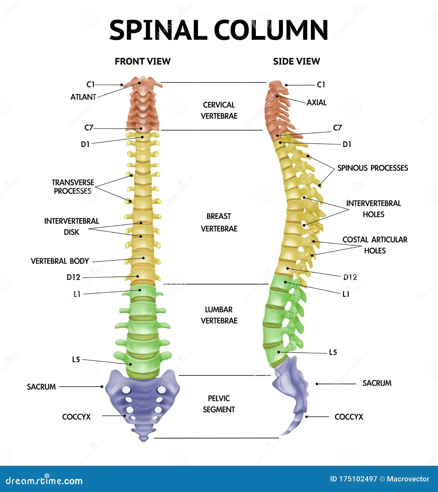
Spinal Manipulation Therapy
Spinal manipulation therapy to the axial skeleton and extraverbal aspects of the spine may serve in the long-term maintenance care of the post-surgical meniscectomy athlete. Chiropractic physicians are spinal practitioners and care deeply about the gliding properties of all components of the axial and appendicular skeleton.
By mobilizing, and if clinically listed, manipulative joints to reduce pain, increase range of motion, and temporarily increase muscle strength and proprioception (6). In a study published in 2010 by the Canadian Chiropractic association researchers hypothesized that spinal manipulation of the lumbar spine would increase muscular strength of the lower extremity via increases in motor neuron excitability and the removal of motor unit inhibition (6). This randomized controlled trial consisted of 50 subjects and discovered no adverse events to spinal manipulation therapy and a 15% strength increase when compared to baseline measurements within the experimental group.
This is of interest to the post-meniscectomy athlete because lower extremity weakness and inhibition of the quadriceps are commonly reported post-surgery.
Review Questions
- The tibiofemoral joint possess how many degrees of freedom
- One
- Two
- Three
- Four
- In regard to arthrokinematics of the tibiofemoral joint, this joint can?
- Flex
- Extend
- Internally Rotate
- Externally Rotate
- All of the Above
- What is the MRI classification used to diagnose meniscal derangement?
- Palmer
- Life
- LaPadre
- National
- What is the name of the surgical procedure used to remove meniscal tissue in order to create a smooth and stable articulating surface of the knee?
- Meniscectomy
- Lateral Release
- Plica Removal
- ACL reconstruction
- What is a clinical concern, as described in this manuscript, for patients immediately following surgical repairment of the meniscus?
- DVT
- Fracture
- Subluxation
- Sprain/Strain
References
- Garcia J R, Ayala S G, Allende F, Mameri E, Haynes M, Familiari F, Geeslin A G, Murrary I, Moatshe G, Verma N N, LaPrade R F. November 2024. Diagnosis and Treatmnet Strategies of Meniscus Root Tears: A Scoping Review. The Orthopedic Journal of Sports Medicine; 12(11): 1-20. Diagnosis and Treatment Strategies of Meniscus Root Tears: A Scoping Review – PubMed
- Tuominen M, Stuart M J, Aubry M, Kannus P, Pakkari J. October 2014. Injuries in Men’s Internation Ice Hockey: A 7-year Study of the International Ice Hockey Federation Adult World Championship Tournaments and Olympic Winter Games. The British Medical Journal; 49(1): 1-7. Injuries in men’s international ice hockey: a 7-year study of the International Ice Hockey Federation Adult World Championship Tournaments and Olympic Winter Games – PubMed
- Meniscal Lesions. Physiopedia. Accessed November 21, 2024. Meniscal Lesions – Physiopedia
- Brunst C, Duerr R, Magnussen R, Flanigan D, Kaeding C. April 2023. Simple Meniscus Repair: Clinical Practice Guidelines. Wexner Medical Center, The Ohio State University: 1-16. Accessed November 21, 2024. OSUWMC-BrandSourceFlierTemplate-Vertical
- Gupta S, Ashish B C, Chavan S K, Gupta P. January 2024. Meniscus Root Tear: Extended Classification and Arthroscopic Repair Techniques. The Journal of Arthoscopy Techniques 13(1): 1-10. Meniscus Root Tear: Extended Classification and Arthroscopic Repair Techniques – PubMed
- Chilibeck P D, Cornish S M, Schulte A, Jantz N, Magnus C R A, Schwanbeck, Juurlink B H J. September 2011. The Effect of Spinal Manipulation on Imbalances in leg Strength. The Journal of the Canadian Chiropractic Association; 55(3): 183-192. The effect of spinal manipulation on imbalances in leg strength – PubMed


Leave a Reply